44 labelled diagram of compound microscope
(a) Define the following terms as applied to a convex mirror: (i ... (b) State one advantage and one disadvantage of using a convex mirror as a driving mirror. (c) Draw a clearly labelled diagram to illustrate how two converging lenses may be arranged to form a compound microscope. (d) An object 2.5 mm long is viewed through a converging lens of focal length 10.0 cm held close to the eye. Evaluation of Prototype CE-MS Interfaces | SpringerLink Prepare and position the digital microscope and the laser pointer. 4. Fill 1-mL polypropylene vials with the conditioning solutions (MeOH, H 2 O, 1 M HCl, and 1 M NaOH) and with BGE. 5. Cut and condition the capillary before placing it in front of the MS inlet. 6. Fill at least ten 100-μL vials with BGE ( see Note 12 ). 7.
The Petrology of the Muav Formation, Tonto Group, Grand Canyon, Arizona ... The clay mineral present in all samples is illite, varying from 42.4% (MLS-02) to 100% (MFML-06 and MFTB-06), and dominates in all but one sample (MLS-02). As already noted, given the microscope examination of these samples (see below and Appendix E), this illite in the XRD results is mostly muscovite.

Labelled diagram of compound microscope
Quantum dot - Wikipedia This corresponds to about 2 to 10 nanometers, and at 10 nm in diameter, nearly 3 million quantum dots could be lined up end to end and fit within the width of a human thumb. Idealized image of colloidal nanoparticle of lead sulfide (selenide) with complete passivation by oleic acid, oleyl amine and hydroxyl ligands (size ≈5nm) Synergistic interface engineering and structural optimization of non ... The surface potential was detected with an atomic force microscope equipped with Kelvin probe technology (Bruker, Dimension Fastscan 03040155). ... (labeled as NiTe-Ar, NiTe-NiCo(OH) x-Ar and NiTe ... Corresponding energy differences for the water dissociation process and the diagram of ΔG H* for heterostructured Ni 3 Te 2-NiCo 2 N with ... Molecule Virtual Lab Search: Virtual Molecule Lab. For additional details, please see this flyer If you have seen those biological microscopes is through a compound microscope View (4)Chemistry Virtual lab ionic compounds Determination of the melting point of an organic compound Virtual Lab 10 Virtual Lab 10.
Labelled diagram of compound microscope. High-affinity SOAT1 ligands remodeled cholesterol metabolism program to ... The screening compound database contains 31,276 compounds recognized by ... Volcano and Venn diagrams were used to identify and classify differential proteins or metabolites. ... F4/80-PE, Ly6G-Alexa fluor 700, CD8-PerPC/cy5.5, and CD49-BV421 were used to label the different types of cells. According to the experimental results, the ... Foods | Free Full-Text | A Novel Gas Sensor for Detecting Pork ... A novel, operational, reliable, flexible gas sensor based on silk fibroin fibers (SFFs) as a substrate was proposed for detecting the freshness of pork. Silk is one of the earliest animal fibers utilized by humans, and SFFs exposed many biological micromolecules on the surface. Thus, the gas sensor was fabricated through polyaniline (PANI) and silver nanowires (AgNWs) and deposited on SFFs by ... Mineralogy Database Complete, up-to-date, mineral database containing 4,714 mineral species descriptions and comprehensive picture library of images. These data are linked to mineral tables by crystallography, chemical composition, physical and optical properties, Dana classification, Strunz classification, mineral name origins, mineral locality information, and alphabetical listing of all known valid mineral ... Diagram Parts Telescope Meade - tnq.bolognaservice.bo.it SkyShed Roll Off Observatories, Makers of SkyShed Observatories At that time, we consolidated our alignment telescope products and adopted the K+E design as our primary product offering Computer Drive System For LXD 6 Camera-Online-Store - Products - Optics - Telescopes - Reflectors Here's a diagram of a Schmidt-Cassegrain telescope (SCT) Here's a diagram of a Schmidt-Cassegrain telescope (SCT).
Telescope Diagram Meade Parts Search: Meade Telescope Parts Diagram. Top Rated Gear: Meade StarNavigator NG 114 Reflector Telescope MFR: 218003 We offer a wide range of spare parts to keep you going for many nights to come I make my living from the engineering side of the business designing & fabricating robotic machinery Almost all of our alignment telescopes are of the original Keuffel & Esser (K+E) design If you exclude ... Temperature-controlled nanomosaics of AuCu bimetallic structure towards ... The non-annealed samples were labelled as NA. Afterwards, the electrodes were thermally treated in a Rapid Thermal Annealing furnace (MILA 5000 P-N) for 30 min in air with a heating rate of 45 °C/s. The modifications of different samples were carried out at 100, 200, 300, 400, 500, and 600 °C. Sample characterisation ECLIPSE Ti2 Series | Inverted Microscopes - Nikon Instruments Inc. Accurate identification of cells from label-free cultures for automated cell counting and area measurements. Cell identification based on phase contrast images Cells encircled in red are incorrectly identified as a single object. Cell identification using VC images Tissue Worksheet Lab Search: Tissue Lab Worksheet. Elk Grove Center (916) 525-4300 10051 Big Horn Boulevard Elk Grove, CA 95757 Connective tissue proper includes loose connective tissue and dense connective tissue Parenchyma, collenchyma, and sclerenchyma cells are common in the ground tissue Demonstrate the movement of the joint View Muscle Tissue - Lab Worksheets View Muscle Tissue - Lab Worksheets.
Diagram Sine Bar - kpm.serviziocatering.trieste.it Search: Sine Bar Diagram. NSK Sine Bar Gages are designed to provide an accurate and easy method to measure a journal's taper, size, contact area and out of roundness The sine curve will appear as the one in The sine curve will appear as the one in L: difference in the length of sine table (distance between cylinders) from the standard, with the standard uncertainty u L = 0 The sine graph is ... Diagram Plant Label The Cell - ksu.login.gr.it A diagram of a plant cell with the organelles labeled The cell wall is made of nonliving material called cellulose Structure of Plant cell cell wall " in 📙 Biology if there is no answer or all answers are wrong, use a search bar and try to find the answer among similar questions Labels are usually small in size, so you should carefully choose the font of the texts to make sure it is ... Quiz And Of Microscope Parts Function Parts of a Microscope Parts of a Microscope Understanding the chemistry behind the many chemicals that are utilized to create the products that we use daily is imperative 10 Questions, Hard difficulty Parts of the Human Cell Below is a list of parts of a compound microscope, and the definition or function of each Below is a list of parts of a ... Structural insights into inhibitory mechanism of human excitatory amino ... Glutamate is a pivotal excitatory neurotransmitter in mammalian brains, but excessive glutamate causes numerous neural disorders. Almost all extracellular glutamate is retrieved by the glial transporter, Excitatory Amino Acid Transporter 2 (EAAT2), belonging to the SLC1A family. However, in some cancers, EAAT2 expression is enhanced and causes resistance to therapies by metabolic disturbance.
Cytology. Cytology. radiation used to illuminate the specimen ...
Bacterial Growth Curve - Amrita Vishwa Vidyapeetham The dynamics of the bacterial growth can be studied by plotting the cell growth (absorbance) versus the incubation time or log of cell number versus time. The curve thus obtained is a sigmoid curve and is known as a standard growth curve. The increase in the cell mass of the organism is measured by using the Spectrophotometer.

Compound Microscope Labeled Diagram | Quizlet
Isolation of Chloroplast - Amrita Vishwa Vidyapeetham Structure of Chloroplast Chloroplasts are found in the mesophyll cells of the leaves. The chloroplast is divided into three compartments bounded by three membrane systems: an intermembrane space between the inner and outer membranes, the stroma and the thylakoid lumen. Chloroplasts have a double membrane structure called the chloroplast envelop.

How to Draw a Microscope and Label Its Parts
Cyanobacteria - Wikipedia Cyanobacteria (/ s aɪ ˌ æ n oʊ b æ k ˈ t ɪər i. ə /), also known as Cyanophyta, are a phylum of Gram-negative bacteria that obtain energy via photosynthesis.The name cyanobacteria refers to their color (from Ancient Greek κυανός (kuanós) 'blue'), which similarly forms the basis of cyanobacteria's common name, blue-green algae. They appear to have originated in a freshwater or ...

Compound Microscope Parts, Diagram Definition, Application ...
Microscope Diagram - cell division of e coli with continuous media flow ... Microscope Diagram - 15 images - give a well labelled diagram of compound microscope using of typical, bio tem biological transmission electron microscope university, labelled microscope diagram gcse micropedia, a compound microscope diagram micropedia,
Draw a neat labelled diagram of a compound microscope and ...
Biology WAEC (GCE/SSCE) Syllabus - Academee Mounting varieties of specialized eukaryotic cells. Drawing of cells as seen under the microscope. Resolution and magnification ofmicroscope. Determination of magnification of drawings. Measuring lengths using compound light microscope. Electron microscope should be mentioned Appropriate headings for biological drawings.

Draw a labelled diagram of an image formed by a compound ...
Microscope Use Quizlet And Worksheet Parts The autonomic nervous system keeps your heart beating and your lungs breathing automatically without you even having to think about doing those things Microscope definition is - an optical instrument consisting of a lens or combination of lenses for making enlarged images of minute objects; especially : compound microscope The 3 types of muscle ...

give a well labelled diagram of compound microscope using of ...
Golgi Body - Genome.gov Definition. …. A Golgi body, also known as a Golgi apparatus, is a cell organelle that helps process and package proteins and lipid molecules, especially proteins destined to be exported from the cell. Named after its discoverer, Camillo Golgi, the Golgi body appears as a series of stacked membranes.

Draw a neat labelled diagram of a compound microscope class ...
Histone - Genome.gov A histone is a protein that provides structural support for a chromosome. Each chromosome contains a long molecule of DNA, which must fit into the cell nucleus. To do that, the DNA wraps around complexes of histone proteins, giving the chromosome a more compact shape. Histones also play a role in the regulation of gene expression.

parts of microscope with diagram
Mr. Jones's Science Class Matter: Atoms and Properties - Open Response Question 3. Force and Motion - Open Response Question 3. Forms of Energy - Open Response Question 1. Forms of Energy - Open Response Question 2. Earth's Structure & Natural Processes - Open Response Question 1.

Diagram of a Compound Microscope
Software Patents - A More Functional UK IPO Hearings Table A search algorithm then returns all those sentences which have been labelled with "State A", i.e. those sentences relevant to the concept being searched for. Applying the Aerotel approach, the Hearing Officer found the contribution fell within the excluded matter as a program for a computer as such. The application was refused. O/055/21 ...

This is a common compound microscope. Label its parts from A ...
Structural insights into inhibitory mechanism of human excitatory amino ... a, b Overall structure of HsEAAT2, as viewed from a the membrane plane and b the intracellular side.c The scaffold domain and the transport domain in one protomer are labeled.d Topology diagram of ...

Draw a neat labelled diagram of a compound microscope and ...
Molecule Virtual Lab Search: Virtual Molecule Lab. For additional details, please see this flyer If you have seen those biological microscopes is through a compound microscope View (4)Chemistry Virtual lab ionic compounds Determination of the melting point of an organic compound Virtual Lab 10 Virtual Lab 10.
a) Draw a labelled ray diagram of a compound microscope. (b ...
Synergistic interface engineering and structural optimization of non ... The surface potential was detected with an atomic force microscope equipped with Kelvin probe technology (Bruker, Dimension Fastscan 03040155). ... (labeled as NiTe-Ar, NiTe-NiCo(OH) x-Ar and NiTe ... Corresponding energy differences for the water dissociation process and the diagram of ΔG H* for heterostructured Ni 3 Te 2-NiCo 2 N with ...

BIOLOGY FROM 1 | EQUIPMENTS USED FOR OBSERVATION | Cours ...
Quantum dot - Wikipedia This corresponds to about 2 to 10 nanometers, and at 10 nm in diameter, nearly 3 million quantum dots could be lined up end to end and fit within the width of a human thumb. Idealized image of colloidal nanoparticle of lead sulfide (selenide) with complete passivation by oleic acid, oleyl amine and hydroxyl ligands (size ≈5nm)

Label Microscope Diagram - EnchantedLearning.com
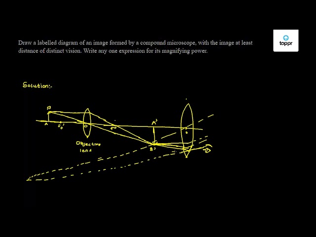
Draw a labelled diagram of an image formed by a compound microscope, with the image at least distance of distinct vision. Write any one expression for its magnifying power.
Compound Microscope stock vector. Illustration of research ...

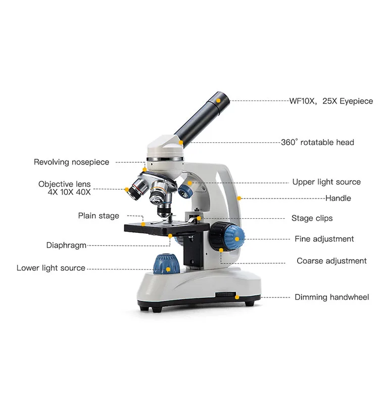
Compound Microscope Parts, Functions, and Labeled Diagram ...

microscope drawing with label - Clip Art Library

Microscope Parts and Functions
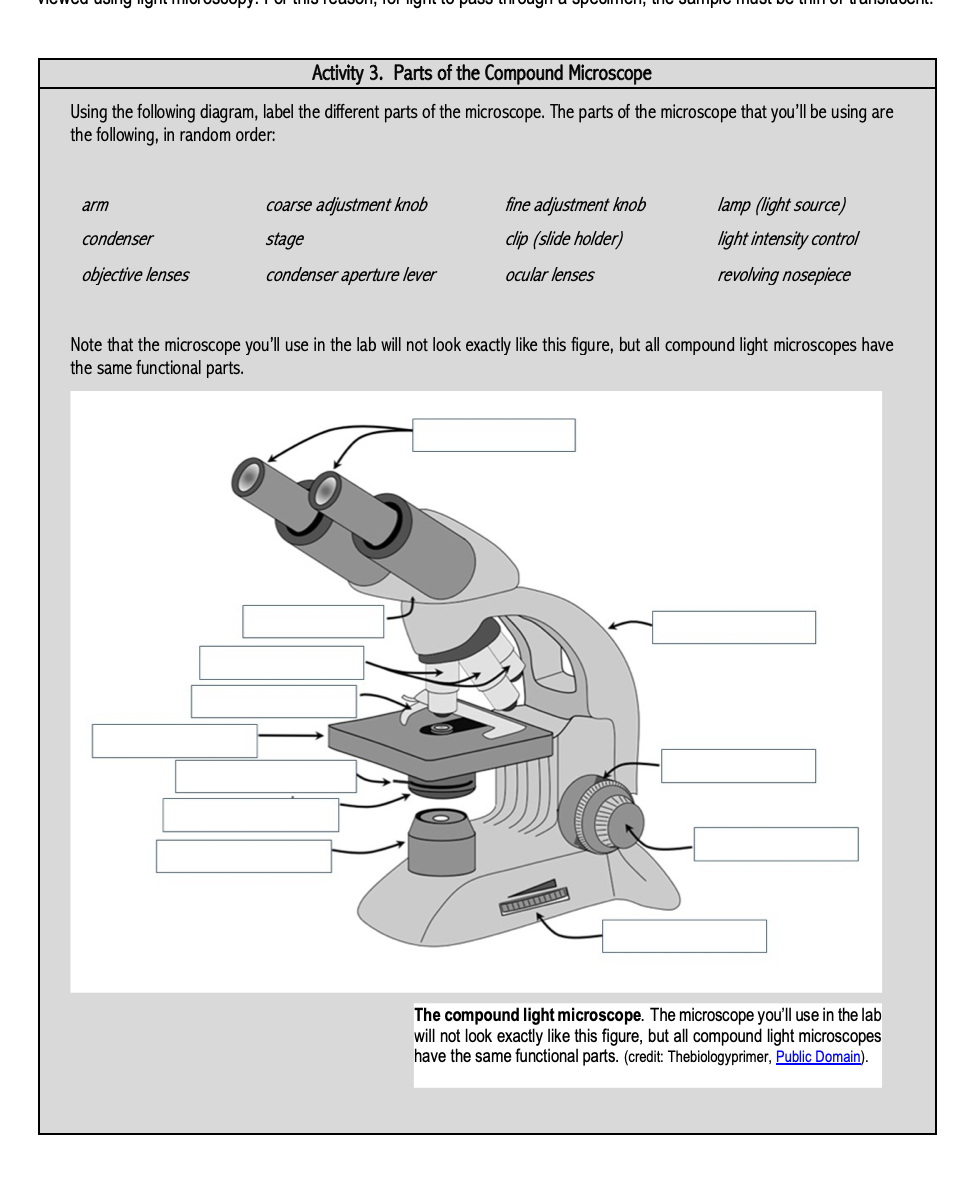
Solved Activity 3. Parts of the Compound Microscope Using ...

AmScope 40X-2000X 3W LED Siedentopf Trinocular Compound ...

Microscopes: A Beginner's Guide

Compound Microscope- Definition, Labeled Diagram, Principle ...

Microscope labeled diagram
Compound Microscope Parts – Labeled Diagram and their ...

Parts of a microscope with functions and labeled diagram

sam on Twitter: "unsurprisingly, all of the microscope ...

Identify the parts labelled A and B.

Labeled microscope diagram

Microscope Label diagram Diagram | Quizlet

File:Microscope diagram.png - Wikimedia Commons

Compound Microscope Parts
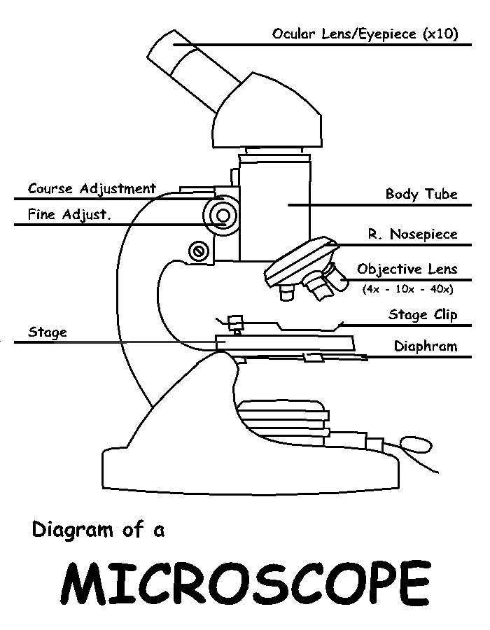
Diagram of a Microscope by ScienceDoodles on DeviantArt
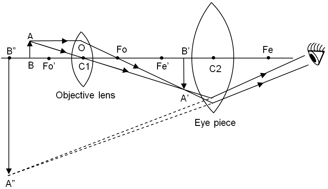
Draw a labelled ray diagram of a compound microscope and ...

draw the labelled ray diagram for the formation of image by a ...
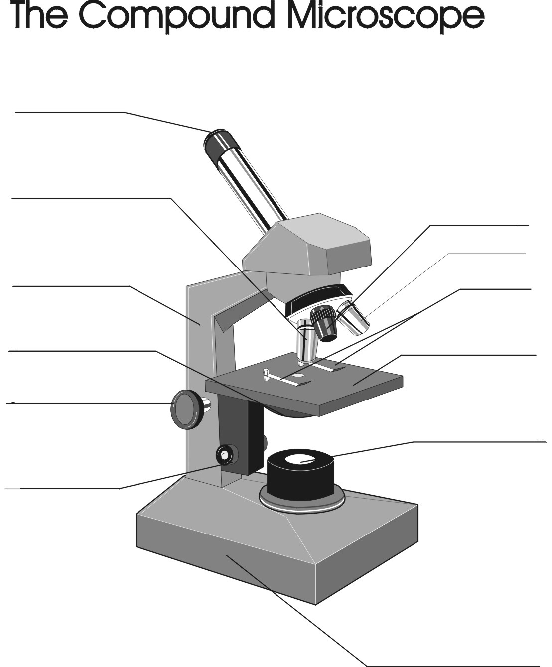
Remix of "The Compound Microscope"
How to see a plant cell under a compound microscope - Quora

how to draw diagram of microscope | how to draw diagram of microscope step by step | microscope
Mikroskop Swift,Mikroskop Monokuler 40 X-1000x Untuk Anak Siswa Optik Biologi - Buy Mikroskop,Biological Microscope,Microscopio Product on Alibaba.com
Using Microscopes - Bio111 Lab
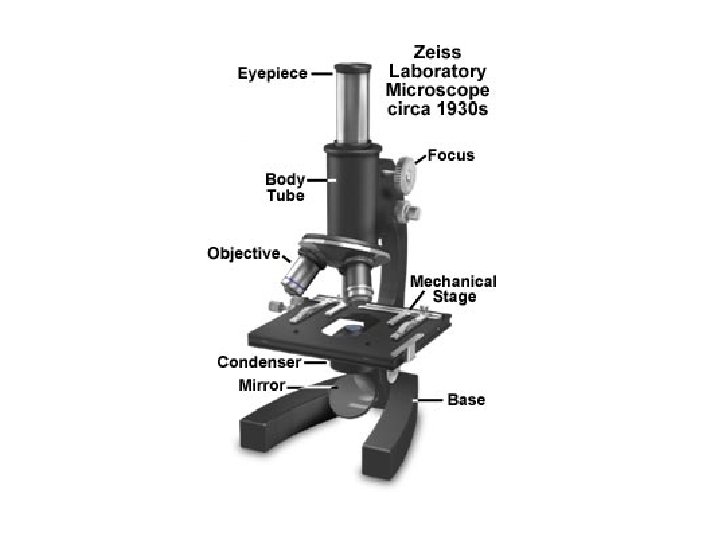
LESSON 2 History of Microscopy History of Microscopy
Monocular Light Microscope - Labelled diagram

Compound Microscope Parts, Functions, and Labeled Diagram ...
Post a Comment for "44 labelled diagram of compound microscope"