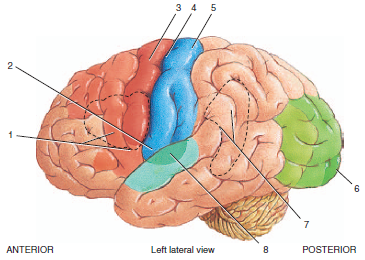
38 correctly label the following functional regions of the cerebral cortex.
Chapter 13 QS Anatomy (Brain and Cranial Nerves) - Quizlet Match the function to the correct lobe of the cerebral cortex. 1. Voluntary skeletal muscle control, verbal communication=frontal lobe 2. Auditory association area=temporal lobe 3. Primary gustatory cortex=insular lobe 4. Somatosensory cortex, somatosensory association area=parietal lobe 5. Primary visual cortex=occipital lobe Unit I Homework Assignment (Chapter 14).docx - 1. Which of... 27. Consider a situation where a stroke or mechanical trauma has occurred resulting in damage to one of the areas of the brain indicated in the image. Drag each label into the proper location in order to identify the area that would most likely have been affected. 28. Correctly label the following functional regions of the cerebral cortex.
Chapter 13 Question Set Flashcards | Quizlet Correctly label the following functional regions of the cerebral cortex. Label the regions involved in interpreting and carrying out speech information. Label the diagram with the terms provided to describe the process of neurulation.
Correctly label the following functional regions of the cerebral cortex.
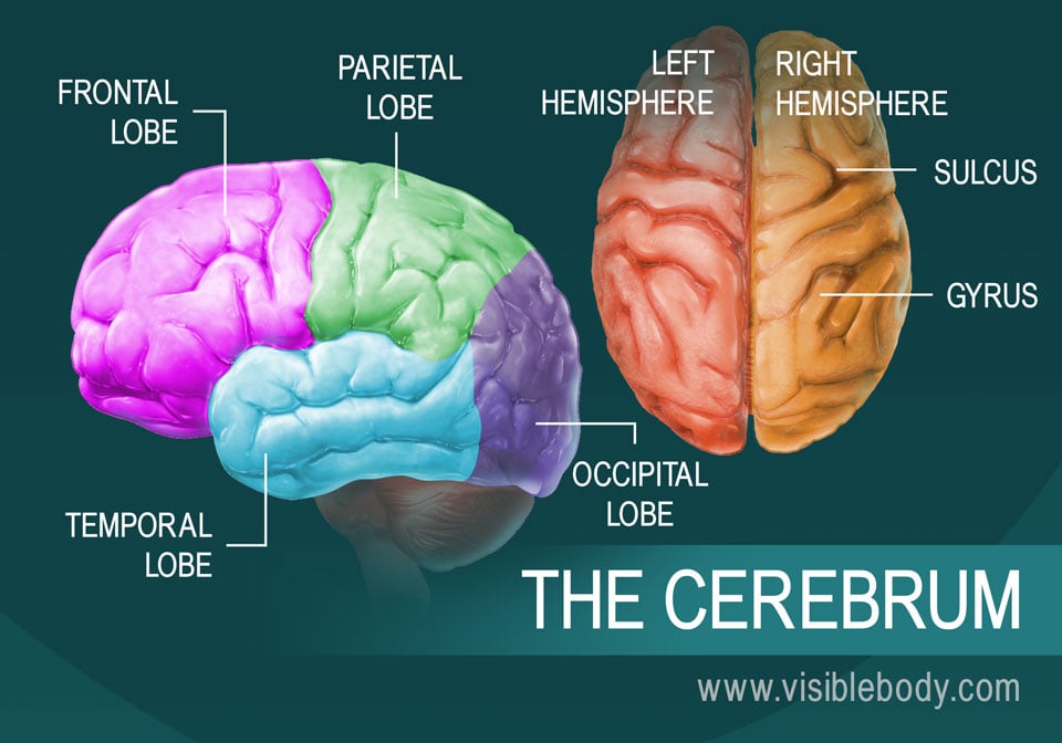
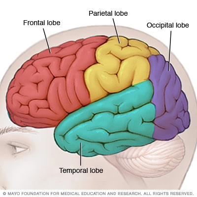
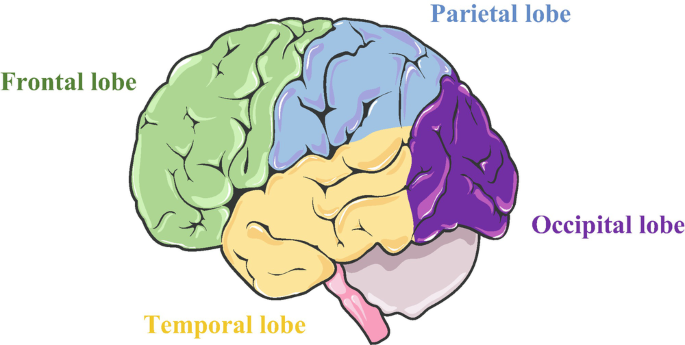
The Four Cerebral Cortex Lobes of the Brain - ThoughtCo Sensory functions interpreted by the cerebral cortex include hearing, touch, and vision. Cognitive functions include thinking, perceiving, and understanding language. Divisions of the Brain Forebrain - encompasses the cerebral cortex and brain lobes. Midbrain - connects the forebrain to the hindbrain. Lobes of the Brain: Cerebral Cortex Anatomy & Function - EZmed The cerebral cortex has 4 main lobes: frontal, parietal, occipital, and temporal. Frontal Lobe The frontal lobe is the largest lobe in the cerebral cortex and is located in the front of the brain as the name suggests. Boundaries There are 2 boundaries that separate the frontal lobe from the adjacent parietal and temporal lobes. Cerebrum: Anatomy, Function, and Treatment - Verywell Health Corpus callosum: A band of white matter that joins the halves of the cerebrum at the deep center of the brain and coordinates nerve signals between each half. Cerebral arteries: Blood vessels that supply the cerebrum with oxygen-rich blood from the heart. There are three cerebral arteries: anterior (front), middle, and posterior (back).; Circle of Willis: A loop of cerebral arteries and other ...
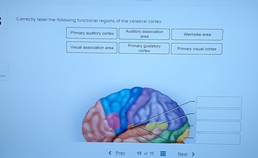
Correctly label the following functional regions of the cerebral cortex.. Exercise-14.docx - The Human Brain 1. Match the letters on... In which of the cerebral lobes are the following functional areas found? auditory cortex: Temporal lobe ... ( n ) 5 tract , whereas one that carries impulses from the cerebrum to lower CNS areas is called a ( n ) 6 tract . The caudate nucleus and putamen are collectively called the 7 . o 1 . ... Label the structures involved with circulation of ... The Functional Areas of the Brain - Human Origin Project The auditory cortex receives input from the ears via lower parts of the auditory system. It then transmits signals back to these areas and other parts of the cerebral cortex. It was subdivided into primary (A1) and secondary (A2), as well as and further association areas. Newer divisions of the auditory cortex are: the core (which includes A1), Solved Correctly label the following functional regions of | Chegg.com Expert Answer. Transcribed image text: Correctly label the following functional regions of the cerebral cortex Auditory association Primary auditory cortex Wernicke area area Primary gustatory cortex Visual association area Primary visual cortex ces Prev 13 of 15 Next. Free Science Flashcards about ANP1040 Exam 4 - StudyStack Correctly label the following functional regions of the cerebral cortex. Postcentral gyrus, Lateral sulcus, Temporal lobe, Precentral gyrus, Central sulcus, Insula, Parietal lobe, Frontal lobe, Occipital lobe: Correctly label the cranial nerves.
Correctly label the following functional regions of the cere +1 (347) 474-1028 info@essayparlour.com Correctly label the following functional regions of the cere Assignment Help biology Correctly label the following functional regions of the cere Format and Features Approximately 275 words/page All paper formats (APA, MLA, Harvard, Chicago/Turabian) Font 12 pt Arial/ Times New Roman Double and single spacing Question : 2.In which of the cerebral lobes the following functional ... 13.Identify the meningeal (or associated) structures described below: 1. outermost meninx covering the brain; composed of tough fibrous connective tissue 2. innermost men... 14.Label the structures involved with circulation of cerebrospinal fluid on the accompanying diagram. Add arrows to the figure above to indicate the flow of cerebrospina... Cerebral cortex: Structure and functions | Kenhub The cerebral cortex (cortex of the brain) is the outer grey matter layer that completely covers the surface of the two cerebral hemispheres. It is about 2 to 4 mm thick and contains an aggregation of nerve cell bodies. This layer is thrown into complex folds, with elevations called gyri and grooves known as sulci. Lab 13/Ch 12 assignments - SET DEFINITIONS FIRST Flashcards - Quizlet 1. Olfaction and hearing are processed in the temporal lobes. 2. The frontal lobe provides critical function in motivation, logical reasoning, expression of emotion, and social attitudes. 3. The occipital lobes house the visual centers, and receive inputs from the optic radiation. 4.
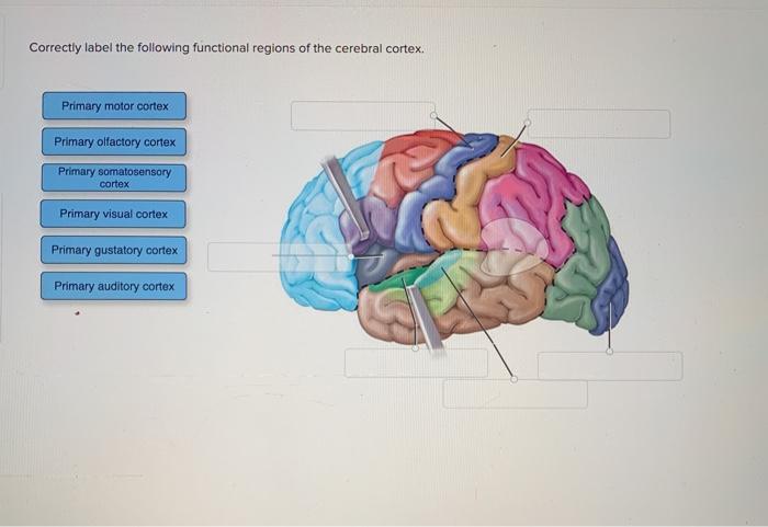
Solved Correctly label the following functional regions of - Chegg Question: Correctly label the following functional regions of the cerebral cortex. Primary motor cortex Primary olfactory cortex Primary somatosensory cortex Primary visual cortex Primary gustatory cortex Primary auditory cortex This problem has been solved! See the answer Show transcribed image text Expert Answer 100% (10 ratings) Cerebral Cortex - Lobes, Fissures, Gyri, and Sulci - GetBodySmart On its lateral and medial surfaces, the cerebral cortex is noticeably convoluted. This increases the surface area of the brain so more nerve cells (or neurons) can be present. The deep furrows are called fissures and shallow ones are called sulci (singluar; sulcus). The ridges between the sulci are known as a gyri (singular; gyrus). Correctly Label The Following Functional Regions Of The Cerebral Cortex ... Correctly Label The Following Functional Regions Of The Cerebral Cortex Somesthetic Association Area Primary Somesthetic Cortex Broca Area Olfactory Association Area Motor Association Area Primary Motor Cortex Prefrontal Cortex Mar 29 2022 10:16 AM Expert's Answer Solution.pdf Next Previous Q: Q: Q: Q: Recent Questions in Basics of Statistics Q: Cerebral Cortex | Facts, Position In Brain, Summary & Function Quick facts: Function: Responsible for thinking and processing information from the five senses. The Cerebral Cortex is made up of tightly packed neurons and is the wrinkly, outermost layer that surrounds the brain. It is also responsible for higher thought processes including speech and decision making . The cortex is divided into four ...
The Human Brain
CBIO Figures Flashcards - Quizlet Correctly label the following functional regions of the cerebral cortex. Image: Correctly label the following functional regions of the cerebral cortex.
Frontal lobe seizures - Symptoms and causes - Mayo Clinic
What midbrain structure is a visual reflex center - Course Hero The parietal lobes exhibit receipt and association of general body sensations. The deepest of the cerebral lobes, the insula is also the smallest and least understood. Cerebral Cortex and Functional Regions Correctly label the following functional regions of the cerebral cortex. Cerebellum: Functions Check all that are functions of the cerebellum.
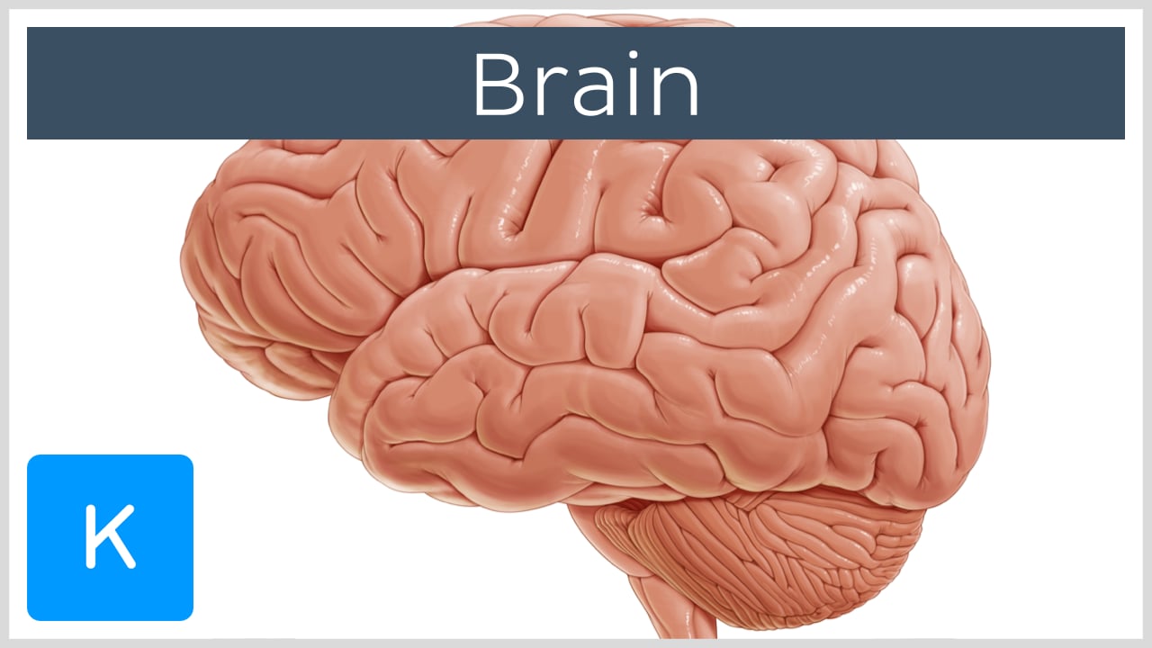
Introduction to the brain
Brodmann areas: Anatomy and functions | Kenhub Synonyms: BA1. Originally defined and numbered into 52 regions by the German anatomist Korbinian Brodmann in the early 1900's, the Brodmann areas of the cerebral cortex are defined by its cytoarchitecture (histological structure and cellular organization). It is important to remember that the same Brodmann area numbers in humans and primates ...
The Human Brain
Cerebral cortex cytoarchitecture and layers | Kenhub Pyramidal cells make up to 75% of the cellular component of the cortex and they are the main output neurons of the cerebral cortex. They vary in size, going from small to gigantic. They usually have one apical dendrite that courses towards the surface of the cortex, and multiple basal dendrites. The number of basal dendrites varies widely, but generally there are more than three to four ...
Solved Correctly label the following functional regions of ...
Cerebral Cortex | Facts, Layers, Levels, Functions & Summary The cerebral cortex can be divided into three basic levels and functions: primary secondary tertiary cortex The hierarchically lowest areas are the primary visual, auditory, somatosensory, and motor cortex. The primary sensory cortex receives information through the thalamus. Primary cortex

Structure and function of language networks in temporal lobe ...
CBIO Figures Flashcards - Quizlet Correctly label the following functional regions of the cerebral cortex. Image: Correctly label the following functional regions of the cerebral cortex.

An atlas of white matter anatomy, its variability, and ...
Solved Correctly label the following functional regions of - Chegg Question: Correctly label the following functional regions of the cerebral cortex Somesthetic association area Primary somesthetic cortex Broca area Olfactory association area Motor association area Primary motor cortex Prefrontal cortex
Solved Correctly label the following functional regions of ...
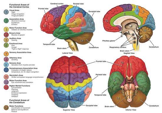
Functional Areas of The Cerebral Cortex - Antranik Functional Areas of The Cerebral Cortex We have two types of functional areas: Sensory areas •Primary Sensory Cortex - makes you aware of a sensation •Association areas - give meaning to/make associations with a sensation •Multimodal Association Areas - make associations between different types of stimuli
.jpg)
Index of /image/Anatomy Brain
Chapter 14 Flashcards - Quizlet Correctly label the following functional regions of the cerebral cortex. ... Image: Correctly label the following meninges of the brain.
Independent and interacting value systems for reward and ...
CBIO Figures Flashcards - Quizlet Correctly label the following functional regions of the cerebral cortex. Image: Correctly label the following functional regions of the cerebral cortex.

CBIO Figures Flashcards | Quizlet
Chapter 14 Worksheet Flashcards | Quizlet Correctly label the following functional regions of the cerebral cortex. Consider a situation where a stroke or mechanical trauma has occurred resulting in damage to one of the areas of the brain indicated in the image. Drag each label into the proper location in order to identify the area that would most likely have been affected.
Mapping brain-wide excitatory projectome of primate ...
CBIO Figures Flashcards | Quizlet Correctly label the following functional regions of the cerebral cortex. Consider a situation where a stroke or mechanical trauma has occurred resulting in damage to one of the areas of the brain indicated in the image. Drag each label into the proper location in order to identify the area that would most likely have been affected.

Cerebrum - an overview | ScienceDirect Topics
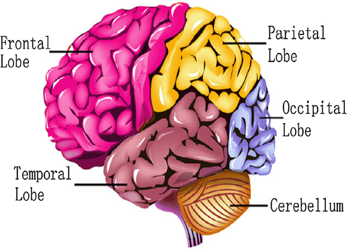
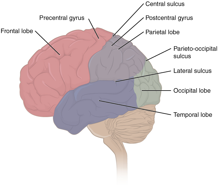
Diagram of the Brain and its Functions - Bodytomy The cerebral cortex is highly convoluted in its structure, which increases the area available for the neurons. The ridges present on the surface of the cortex are called gyri, whereas the grooves or fissures separating them are called sulci. ... The temporal lobe is a region of the cerebral cortex that is present beneath the Sylvian fissure. It ...
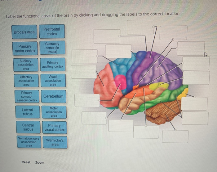
Solved Label the functional areas of the brain by clicking ...
A. the stroke caused damage to Joe's frontal eye field which interfered with his effort to touch his chin. B. the stroke caused damage to Joe's right primary motor cortex. C. the stroke caused damage to Joe's left premotor cortex. D. based on the doctor's observations, none of the listed answers are correct conclusions.

Lateralization of brain function - Wikipedia
Cerebrum: Anatomy, Function, and Treatment - Verywell Health Corpus callosum: A band of white matter that joins the halves of the cerebrum at the deep center of the brain and coordinates nerve signals between each half. Cerebral arteries: Blood vessels that supply the cerebrum with oxygen-rich blood from the heart. There are three cerebral arteries: anterior (front), middle, and posterior (back).; Circle of Willis: A loop of cerebral arteries and other ...
Biomaterials Developments for Brain Tissue Engineering ...
Lobes of the Brain: Cerebral Cortex Anatomy & Function - EZmed The cerebral cortex has 4 main lobes: frontal, parietal, occipital, and temporal. Frontal Lobe The frontal lobe is the largest lobe in the cerebral cortex and is located in the front of the brain as the name suggests. Boundaries There are 2 boundaries that separate the frontal lobe from the adjacent parietal and temporal lobes.
Cerebral Cortex - Physiopedia
The Four Cerebral Cortex Lobes of the Brain - ThoughtCo Sensory functions interpreted by the cerebral cortex include hearing, touch, and vision. Cognitive functions include thinking, perceiving, and understanding language. Divisions of the Brain Forebrain - encompasses the cerebral cortex and brain lobes. Midbrain - connects the forebrain to the hindbrain.

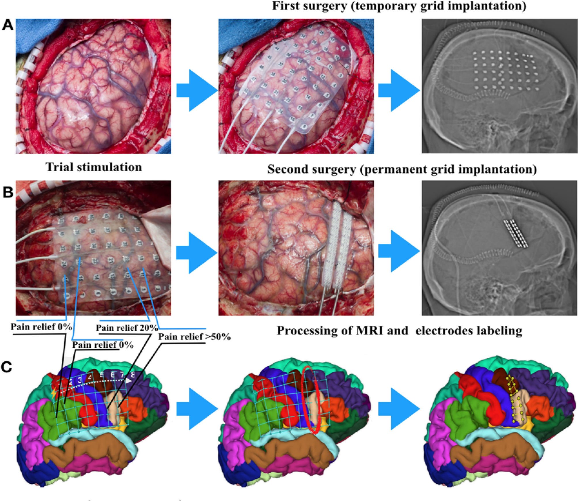
The utility of arterial spin labeling in the presurgical ...

CBIO Figures Flashcards | Quizlet
Untitled
Human emotion recognition from EEG-based brain–computer ...

Functional Relevance of Endocannabinoid-Dependent Synaptic ...
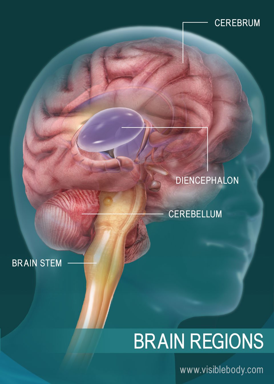
The Human Brain

Connectivity‐based parcellation of normal and anatomically ...
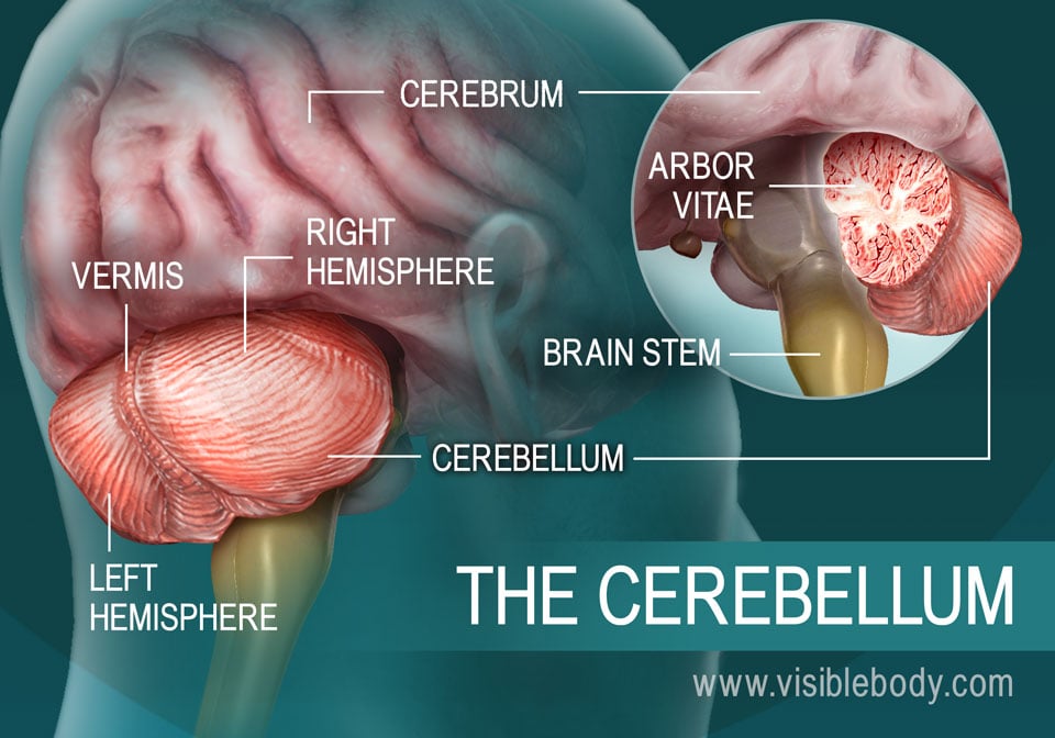
Brainstem: Definition, anatomy, parts, function | Kenhub

An atlas of white matter anatomy, its variability, and ...

AHCDW10Notes82.pdf - 82. Award: 10.00 points Problems? Adjust ...
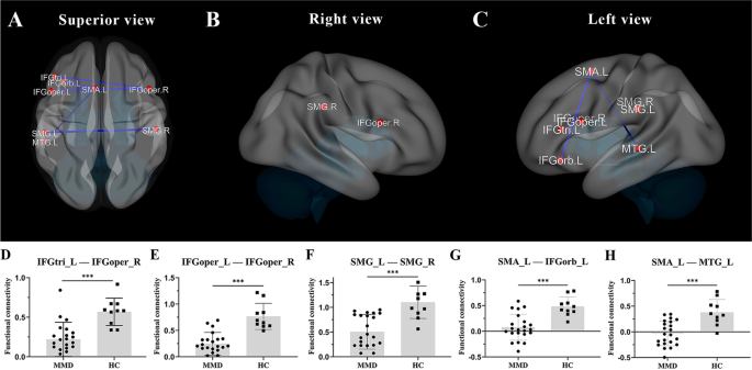
Abnormal brain functional and structural connectivity between ...
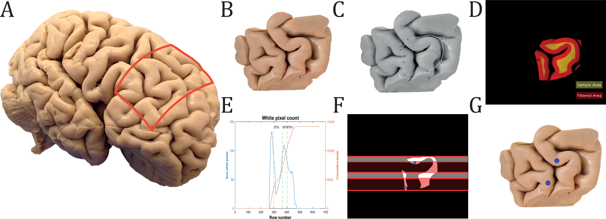
Cellular 3D-reconstruction and analysis in the human cerebral ...

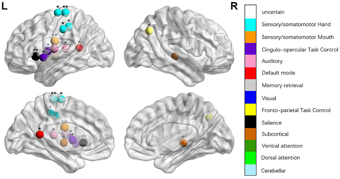
Description of the brain functional regions and their ...
Cerebral Cortex: Anatomy | Concise Medical Knowledge

Solved Chapter 14 Homework Interactive Questi... Saved Help ...
motor and movement problems in children
:max_bytes(150000):strip_icc()/cerebral_cortex-56a09b863df78cafdaa3301d.jpg)
The Four Cerebral Cortex Lobes of the Brain
Solved] Which of the following functional regions of the ...
Frontiers | Disrupted Brain Connectivity Networks in Aphasia ...
Pre-motor versus motor cerebral cortex neuromodulation for ...
Anatomy Exam 2 Flashcards - Easy Notecards
Post a Comment for "38 correctly label the following functional regions of the cerebral cortex."