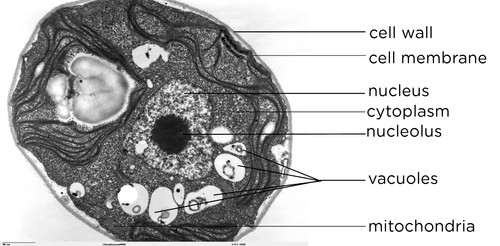
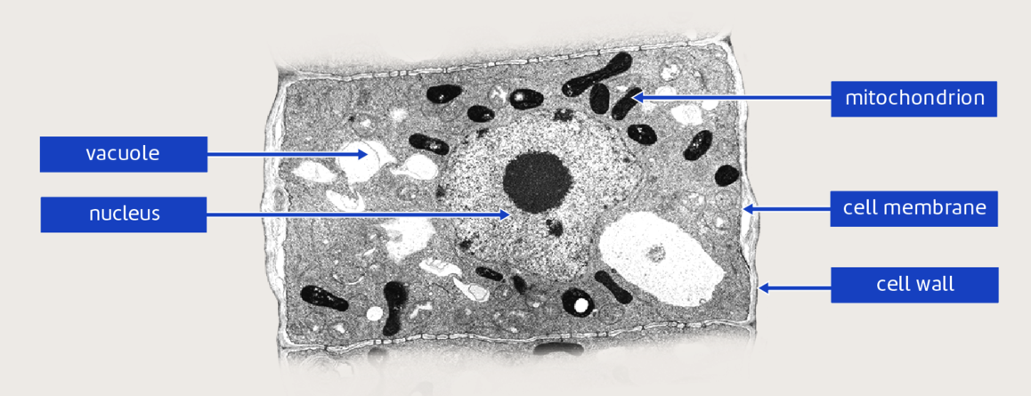
44 nucleus electron micrograph labelled
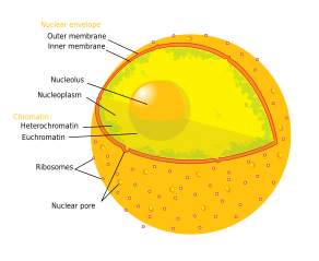
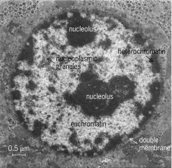
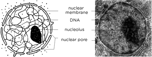
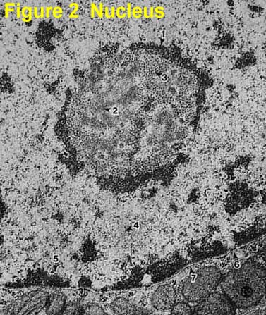
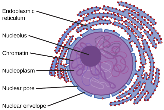
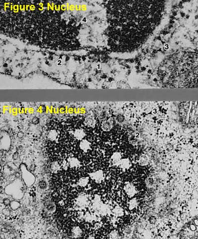
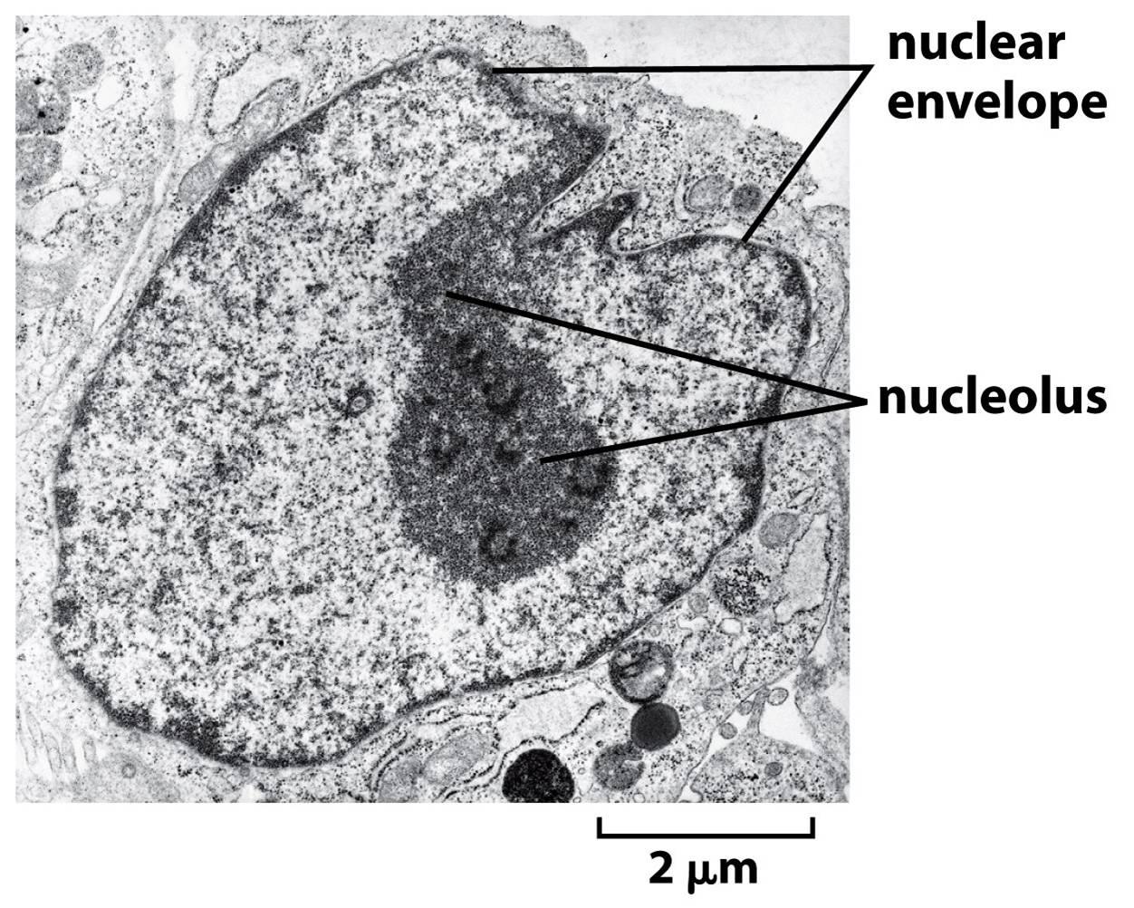
Electron Micrographs Figure 1 Micrograph of a nucleus. 1. Heterochromatin: 2. Euchromatin: 3. Nucleolus: 4. Nucleolar associated chromatin: 5. Nuclear envelope. Figure 2 Micrograph ... Cell Biology - Buffalo Public Schools microscope was invented – the electron microscope. ... nucleus and membrane-bound cellular organelles. The ... Write on or label the final drawing in.
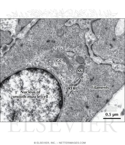
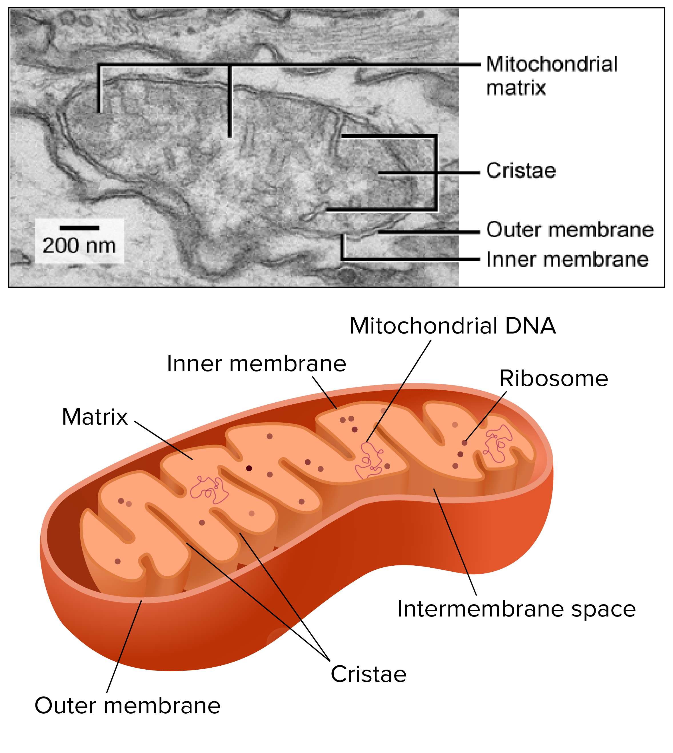
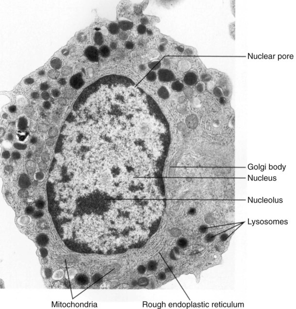
Electron Micrographs of Cell Organelles | Zoology The Electron Micrograph of Mitochondria: ... (1) The name mitochondria was given by Benda (1898) and their ma n function was brought to light by Kingsbury (1912).
Nucleus electron micrograph labelled
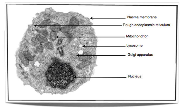
Cell Micrographs | BioNinja A micrograph is a photo or digital image taken through a microscope to show a magnified image of a specimen. While organelles have identifying structures, ...
Nucleus electron micrograph labelled. Cell Micrographs | BioNinja A micrograph is a photo or digital image taken through a microscope to show a magnified image of a specimen. While organelles have identifying structures, ...
Electron Micrograph of Actin and Intermediate Filaments In ...

BIOL 230 Lecture Guide - Eukaryotic Cell
Mitochondria and chloroplasts (article) | Khan Academy
Nucleolus - Wikipedia

587 Transmission Electron Micrograph Images, Stock Photos ...

An electron micrograph of control mouse showing normal ...

Cellular organelles | Deranged Physiology
A tour of the cell: View as single page
Cell nucleus | Article about Cell nucleus by The Free Dictionary
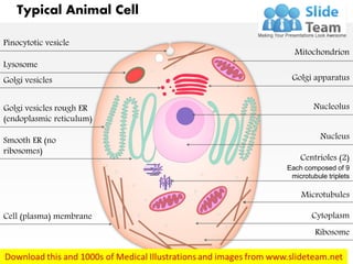
A typical animal cell (as seen in an electron microscope ...

Animal & Plant Cells (1.2.1) | CIE A Level Biology Revision ...

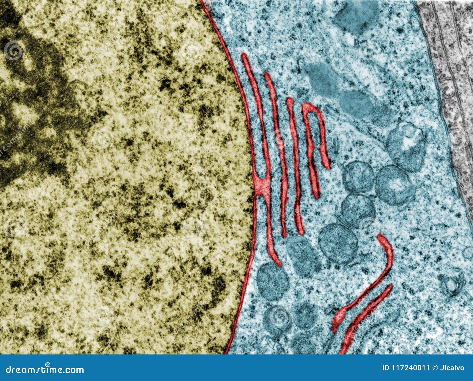
Figure, Transmission Electron Micrograph of Rough Endoplasmic ...
Cellular Structure and Function | Oncohema Key

File:Anatomy and physiology of animals animal cell electron ...

The Nucleus - Cell Organelles Ep 1 - Zoë Huggett Tutorials
1.1 Cell structure | Cells as the basic units of life | Siyavula

An electron micrograph of a barley nucleus including a ...

1,845 Electron Micrograph Images, Stock Photos & Vectors ...

The transmission electronmicroscope shows parts of two ...
Natural Sciences Grade 9

Cell Micrographs | BioNinja
Electron Micrographs
The Cell: The Histology Guide
Draw a neat and labelled diagram of Nucleus. - Sarthaks ...

Nucleus. tem. False colour transmission electron microscope ...
3.3 Cells under the microscope - Stile

Transmission electron micrograph of an animal cell - Stock ...

GCE CIE Biology - Animal and Plant Cell Structures and ...

plant cell label electron micrograph Diagram | Quizlet
3.3 Eukaryotic Cells – Concepts of Biology – 1st Canadian Edition
Electron Micrographs

DP Topic 1.1 / 1.2 | Biology - Quizizz
9700 QR Dynamic Papers Biology al Cambridge
cell and organelles Dr.Jastrow's electron microscopic atlas
Nuclear envelope. TEM stock image. Image of micrograph ...

Draw a diagram to show the structure of a neuron with ...

Electron Micrograph of Plasma Cells In Connective Tissue
Chapter 4: DNA, Chromosomes, and Genomes Flashcards | Chegg.com
SOLVED: Name the structure at the blue arrow in the electron ...

Cytology | Veterian Key
IJMS | Free Full-Text | Visualization of Chromatin in the ...
Electron Micrographs

1.2 Skill: Interpretation of electron micrographs
IB Biology Notes - 2.3 Eukaryotic cells
Post a Comment for "44 nucleus electron micrograph labelled"