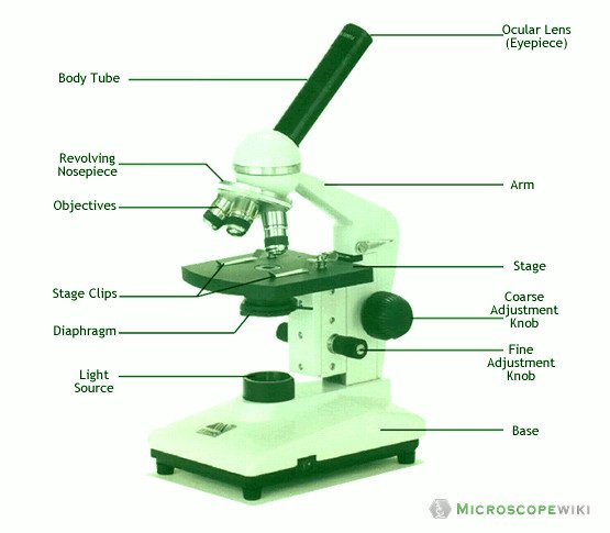
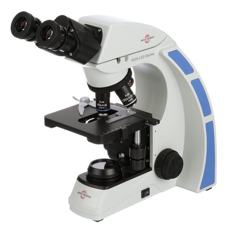
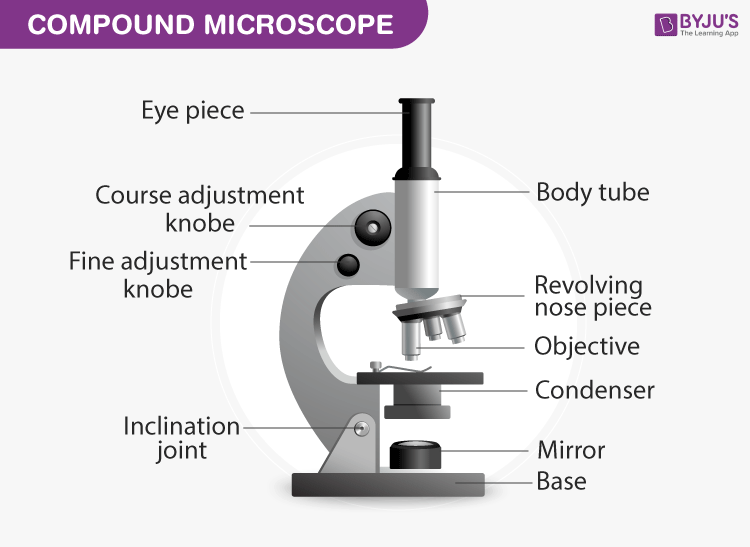
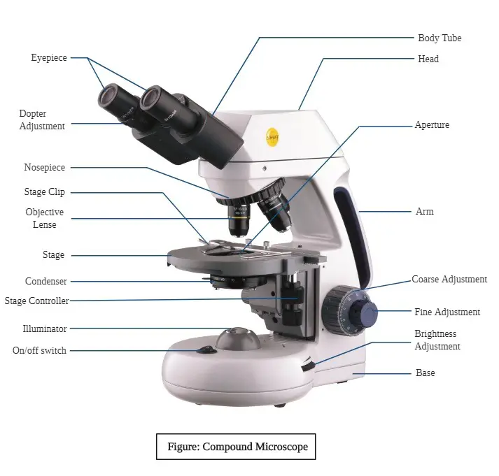
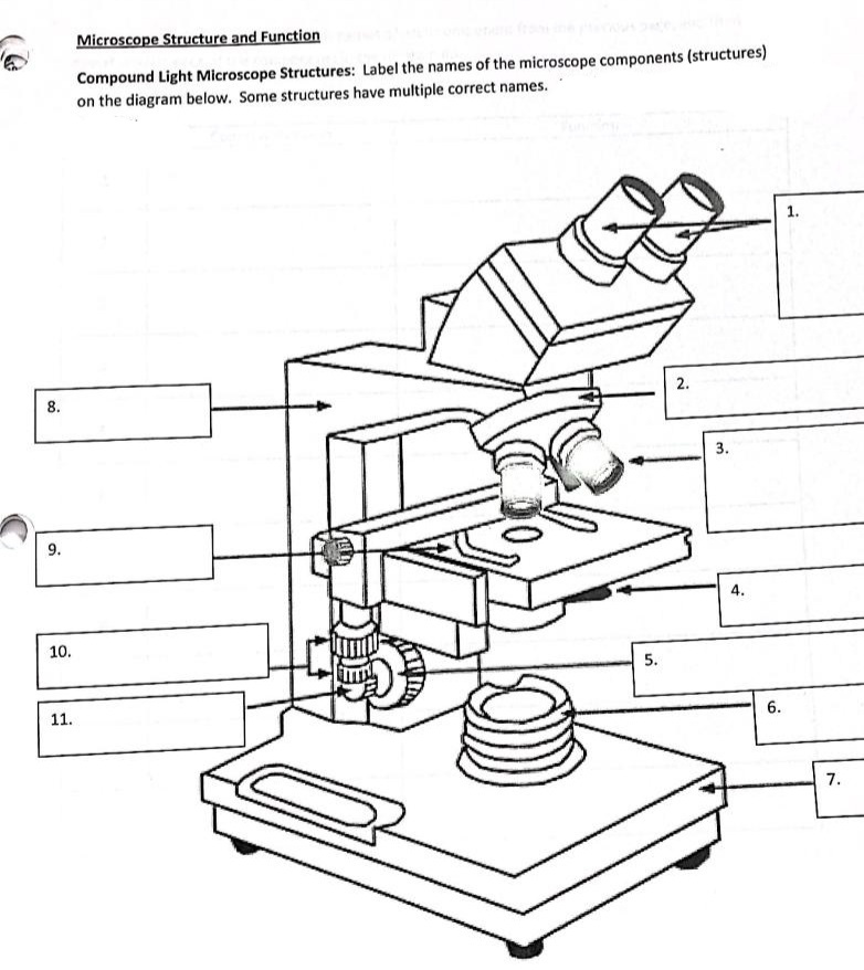
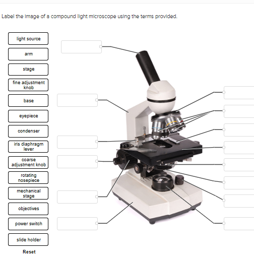
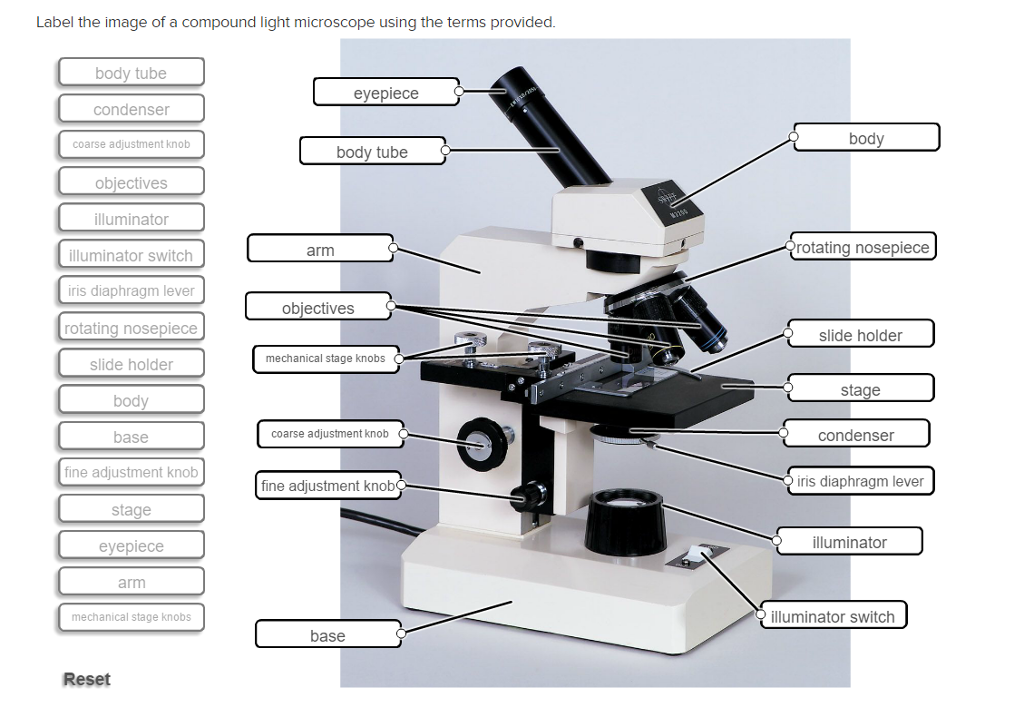
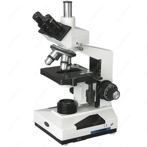
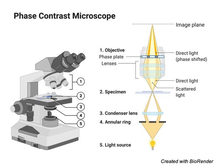
42 label the compound light microscope
› light-microscopesCompound Light Microscopes | Products | Leica Microsystems Aug 08, 2018 · A compound microscope uses optics to produce a magnified image of a sample so that with details of it can be observed that are undetectable with the naked eye. The most basic optics of a compound microscope has at least 2 lenses: i) an objective placed nearby the sample which creates a magnified, real image of it and ii) eyepieces or oculars ... › anton-van-leeuwenhoek-1991633Antonie van Leeuwenhoek, Father of Microbiology - ThoughtCo Jul 21, 2019 · Also credited with the invention of the microscope about the same time was Hans Lippershey, the inventor of the telescope. Their work led to others' research and development on telescopes and the modern compound microscope, such as Galileo Galilei, Italian astronomer, physicist, and engineer whose invention was the first given the name ...
Compound Light Microscope Labelling Quiz - PurposeGames.com This is an online quiz called Compound Light Microscope Labelling There is a printable worksheet available for download here so you can take the quiz with pen and paper. Your Skills & Rank Total Points 0 Get started! Today's Rank -- 0 Today 's Points One of us! Game Points 15 You need to get 100% to score the 15 points available Actions

Label the compound light microscope
Compound Microscope: Definition, Diagram, Parts, Uses, Working ... - BYJUS The parts of a compound microscope can be classified into two: Non-optical parts Optical parts Non-optical parts Base The base is also known as the foot which is either U or horseshoe-shaped. It is a metallic structure that supports the entire microscope. Pillar The connection between the base and the arm are possible through the pillar. Arm Parts of a microscope with functions and labeled diagram 19.04.2022 · Q. Differentiate between a condenser and an Abbe condenser. Ans. Condensers are lenses that are used to collect and focus light from the illuminator into the specimen. They are found under the stage next to the diaphragm of the microscope. They play a major role in ensuring clear sharp images are produced with a high magnification of 400X and above. Fluorescence Microscopy - Explanation and Labelled Images 16.12.2020 · A fluorescence microscope uses a higher intensity light to illuminate the samples. Parts of a Fluorescence Microscope A powerful light source (xenon or mercury arch lamp) : The light emitted from the mercury arc lamp is 10-100 times brighter than most incandescent lamps and provides light in a wide range of wavelengths, from ultra-violet to the infrared.
Label the compound light microscope. Parts of a Compound Light Microscope - YouTube Made specifically for students of Mr Piper to learn the basic about the parts of a microscope. Definition and Examples of Compound-Complex Sentences 24.07.2019 · How, Why, and When to Use Compound-Complex Sentences "The compound-complex sentence consists of two or more independent clauses and one or more dependent clauses. This syntactic shape is essential in representing complex relationships and so is frequently put to use in various forms of analytical writing, especially in academic writing.It is … Label the microscope — Science Learning Hub Drag and drop the text labels onto the microscope diagram. If you want to redo an answer, click on the box and the answer will go back to the top so you can move it to another box. If you want to check your answers, use the Reset incorrect button. This will reset incorrect answers only. PDF Parts of the Light Microscope - Science Spot Parts of the Light Microscope EYEPIECEContains the OCULAR lens NOSEPIECEHolds the HIGH- and LOW-objective LENSES; can bechange MAGNIFICATION. OBJECTIVE LENSESMagnification ranges from 10 X to 40 X STAGE CLIPSHOLD the slide in place STAGESupports the SLIDE being viewed K. ARMUsed to SUPPORT the microscope when carried COARSE ADJUSTMENT KNOB
Compound Microscope Parts The three basic, structural components of a compound microscope are the head, base and arm. Head/Body houses the optical parts in the upper part of the microscope Base of the microscope supports the microscope and houses the illuminator Arm connects to the base and supports the microscope head. It is also used to carry the microscope. UD Virtual Compound Microscope - University of Delaware ©University of Delaware. This work is licensed under a Creative Commons Attribution-NonCommercial-NoDerivs 2.5 License.Creative Commons Attribution-NonCommercial-NoDerivs 2.5 … Parts of Stereo Microscope (Dissecting microscope) – labeled … Compared to a compound microscope where the objectives attached to the nosepiece can be seen and identified individually (based on color bands and their respective labels), the objectives of a dissecting microscope are located in a cylindrical cone and, therefore, are not directly seen. For the stereo microscope that comes with multiple objective lens sets (fixed power style), the … Microscopy Pre-lab Activities - University of Delaware Microscope controls: turn knobs (click and hold on upper or lower portion of knob) throw switches (click and drag) turn dials (click and drag) move levers (click and drag) changes lenses (click and drag on objective housing) select a specimen (click on a slide) adjust oculars (in "through" view, with the light on, click and drag to move oculars closer or further apart) Designed and …
Microscope, Microscope Parts, Labeled Diagram, and Functions 19.01.2022 · Revolving Nosepiece or Turret: Turret is the part of the microscope that holds two or multiple objective lenses and helps to rotate objective lenses and also helps to easily change power. Objective Lenses: Three are 3 or 4 objective lenses on a microscope. The objective lenses almost always consist of 4x, 10x, 40x and 100x powers. The most common eyepiece … Antonie van Leeuwenhoek, Father of Microbiology - ThoughtCo 21.07.2019 · Their work led to others' research and development on telescopes and the modern compound microscope, such as Galileo Galilei, Italian astronomer, physicist, and engineer whose invention was the first given the name "microscope." The compound microscopes of Leeuwenhoek's time had issues with blurry figures and distortions and could magnify only up to … www1.udel.edu › biology › ketchamMicroscopy Pre-lab Activities - University of Delaware Microscope controls: turn knobs (click and hold on upper or lower portion of knob) throw switches (click and drag) turn dials (click and drag) move levers (click and drag) changes lenses (click and drag on objective housing) select a specimen (click on a slide) Compound Microscope Labeled Diagram | Quizlet QUESTION. The total magnification of a specimen being viewed with a 10X ocular lens and a 40X objective lens is. 15 answers. QUESTION. a mosquito beats its wings up and down 600 times per second, which you hear as a very annoying 600 Hz sound. if the air outside is 20 C, how far would a sound wave travel between wing beats. 2 answers.
Parts of a microscope with functions and labeled diagram - Microbe Notes Microscopic illuminator - This is the microscopes light source, located at the base. It is used instead of a mirror. It captures light from an external source of a low voltage of about 100v. Condenser - These are lenses that are used to collect and focus light from the illuminator into the specimen.
Compound Microscope Parts - Labeled Diagram and their Functions A compound microscope is the most common type of light (optical) microscopes. The term "compound" refers to the microscope having more than one lens. Basically, compound microscopes generate magnified images through an aligned pair of the objective lens and the ocular lens.
Compound Microscope - Diagram (Parts labelled), Principle and Uses As the name suggests, a compound microscope uses a combination of lenses coupled with an artificial light source to magnify an object at various zoom levels to study the object. A compound microscope: Is used to view samples that are not visible to the naked eye Uses two types of lenses - Objective and ocular lenses
Microscope Labeling - The Biology Corner Microscope Labeling. Shannan Muskopf May 31, 2018. This simple worksheet pairs with a lesson on the light microscope, where beginning biology students learn the parts of the light microscope and the steps needed to focus a slide under high power. The labeling worksheet could be used as a quiz or as part of direct instruction where students ...
Compound Light Microscope | Science Quiz - Quizizz 12 Questions Show answers. Supports the stage and upper part of the microscope; contains focus adjustment knobs. Wheel or lever that adjusts the amount of light that passes through the hole in the stage and into the specimen. The part of the microscope through which you look; contains usually 10x lens. Raises and lowers the stage or objective ...
Compound Microscope- Definition, Labeled Diagram, Principle, Parts, Uses Alternatively, the magnification of the compound microscope is given by: m = D/ fo * L/fe where, D = Least distance of distinct vision (25 cm) L = Length of the microscope tube fo = Focal length of the objective lens fe = Focal length of the eye-piece lens Parts of a Compound Microscope Eyepiece And Body Tube.
Solved Label the image of a compound light microscope using - Chegg Expert Answer. 100% (17 ratings) Transcribed image text: Label the image of a compound light microscope using the terms provided.
Labelled Diagram of Compound Microscope The below mentioned article provides a labelled diagram of compound microscope. Part # 1. The Stand: The stand is made up of a heavy foot which carries a curved inclinable limb or arm bearing the body tube. The foot is generally horse shoe-shaped structure (Fig. 2) which rests on table top or any other surface on which the microscope in kept.
Bright-field microscope (Compound light microscope) - Diagram (Parts ... Bright-field Microscope. A bright-field microscope, also known as a compound light microscope is among the simplest of optical microscopes. Optical microscopes employ visible light and a series of lenses to magnify the specimen and view it in detail. A bright-field microscope uses light rays to create a dark image against a bright background ...
Microscope Labeling Game - PurposeGames.com About this Quiz. This is an online quiz called Microscope Labeling Game. There is a printable worksheet available for download here so you can take the quiz with pen and paper. This quiz has tags. Click on the tags below to find other quizzes on the same subject. Science.
Compound Light Microscopes | Products | Leica Microsystems 08.08.2018 · The maximum useful magnification value achieved with any type of light microscope depends on its ultimate resolving power or maximal resolution. The resolution depends on the microscope objective lens numerical aperture (NA). At low magnification values, the NA is small leading to a low resolution. At high magnification values, the NA is high ...
› p-3470-what-is-aWhat is a Compound Microscope? | Microscope World Blog A compound microscope is a high power (high magnification) microscope that uses a compound lens system. A compound microscope has multiple lenses: the objective lens (typically 4x, 10x, 40x or 100x) is compounded (multiplied) by the eyepiece lens (typically 10x) to obtain a high magnification of 40x, 100x, 400x and 1000x.
Parts of a Compound Microscope (And their Functions) - Scope Detective A compound microscope is the most common microscope you can get and the type you'll typically see in a lab or hobbyist's study. These microscopes tend to have. ... For a compound light microscope, there are usually two sets of ocular lenses in a purchase: 10X and 20X. However, this isn't always the case, particularly with Barlow lenses ...
Compound Light Microscope Optics, Magnification and Uses Magnification. In order to ascertain the total magnification when viewing an image with a compound light microscope, take the power of the objective lens which is at 4x, 10x or 40x and multiply it by the power of the eyepiece which is typically 10x. Therefore, a 10x eyepiece used with a 40X objective lens, will produce a magnification of 400X.
www1.udel.edu › biology › ketchamUD Virtual Compound Microscope - University of Delaware ©University of Delaware. This work is licensed under a Creative Commons Attribution-NonCommercial-NoDerivs 2.5 License.Creative Commons Attribution-NonCommercial-NoDerivs 2
Compound Microscope Parts, Functions, and Labeled Diagram The total magnification of a compound microscope is calculated by multiplying the objective lens magnification by the eyepiece magnification level. So, a compound microscope with a 10x eyepiece magnification looking through the 40x objective lens has a total magnification of 400x (10 x 40).
Parts of the Microscope with Labeling (also Free Printouts) Parts of the Microscope with Labeling (also Free Printouts) By Editorial Team March 7, 2022 A microscope is one of the invaluable tools in the laboratory setting. It is used to observe things that cannot be seen by the naked eye. Table of Contents 1. Eyepiece 2. Body tube/Head 3. Turret/Nose piece 4. Objective lenses 5. Knobs (fine and coarse) 6.
Compound Microscope: Parts of Compound Microscope - BYJUS (A) Mechanical Parts of a Compound Microscope 1. Foot or base It is a U-shaped structure and supports the entire weight of the compound microscope. 2. Pillar It is a vertical projection. This stands by resting on the base and supports the stage. 3. Arm The entire microscope is handled by a strong and curved structure known as the arm. 4. Stage
› compound-complex-sentenceDefinition and Examples of Compound-Complex Sentences - ThoughtCo Jul 24, 2019 · This is not to say that the compound-complex sentence invites confusion: on the contrary, when handled carefully, it has the opposite effect—it clarifies the complexity and enables readers to see it clearly." (David Rosenwasser and Jill Stephen, Writing Analytically, 6th ed. Wadsworth, 2012) "Compound-complex sentences get unwieldy in a hurry ...
What is a Compound Microscope? | Microscope World Blog A metallurgical microscope is a compound microscope that may have transmitted and reflected light, or just reflected light. This reflected light shines down through the objective lens. Metallurgical compound microscopes are specifically used in industrial settings to view samples at high magnification (such as metals) that will not allow light to pass through them. …
Microscope Parts and Functions Objective lenses: One of the most important parts of a compound microscope, as they are the lenses closest to the specimen. A standard microscope has three, four, or five objective lenses that range in power from 4X to 100X.
label parts of a compound microscope - TeachersPayTeachers This is a set of 3 tiered readings. Students will read a passage about the how to use a compound light microscope. Students will use textual evidence to answer questions and label the different parts of the microscope. It also allows students to gain prior knowledge about the compound microscope. Version A provides the most support for students.
Compound Light Microscope: Everything You Need to Know A compound light microscope is a type of light microscope that uses a compound lens system, meaning, it operates through two sets of lenses to magnify the image of a specimen. It's an upright microscope that produces a two-dimensional image and has a higher magnification than a stereoscopic microscope.
Solved Part C Identify the parts of the compound light - Chegg Transcribed image text: Part C Identify the parts of the compound light microscope. Drag the appropriate label to the appropriate target. Reset C Ocular lens Arm Revolving nosepiece Condenser focus knob Objective lens Stage Condenser Light intensity dial Light source Base Condenser aperture diaphragm Fine focusing knob Coarse focusing knob lai.
A. Microscope & Cells (1).docx - Label the Compound Light... A cell biologist would choose an electron microscope rather than a light microscope because an electron microscope is capable of magnifications of up to 1 500 000 x… research on cells has led to major breakthroughs in medicine and industry due to the electron microscope . Today , compound light microscope can only permit to a level of ...
microscopeinternational.com › fluorescence-microscopyExplanation and Labelled Images - New York Microscope Company Dec 16, 2020 · A fluorescence microscope uses a higher intensity light to illuminate the samples. Parts of a Fluorescence Microscope A powerful light source (xenon or mercury arch lamp) : The light emitted from the mercury arc lamp is 10-100 times brighter than most incandescent lamps and provides light in a wide range of wavelengths, from ultra-violet to the ...
Name the Parts of the Compound Light Microscope - Sporcle Name the Parts of the Compound Light Microscope Quiz - By KellyHarrison. Science microscope. QUIZ LAB SUBMISSION. Random Science or microscope Quiz.
label a microscope worksheet Compound Light Microscope Labeled - Made By Creative Label labels-creative.com. microscope compound light parts labeled label worksheet labels source homeschooldressage creative does aim ppt. Structure Of A Leaf With Diagram And Detail Explaination Plz Its Urgent . stomata layer explaination epidermis cuticle urgent
Fluorescence Microscopy - Explanation and Labelled Images 16.12.2020 · A fluorescence microscope uses a higher intensity light to illuminate the samples. Parts of a Fluorescence Microscope A powerful light source (xenon or mercury arch lamp) : The light emitted from the mercury arc lamp is 10-100 times brighter than most incandescent lamps and provides light in a wide range of wavelengths, from ultra-violet to the infrared.
Parts of a microscope with functions and labeled diagram 19.04.2022 · Q. Differentiate between a condenser and an Abbe condenser. Ans. Condensers are lenses that are used to collect and focus light from the illuminator into the specimen. They are found under the stage next to the diaphragm of the microscope. They play a major role in ensuring clear sharp images are produced with a high magnification of 400X and above.
Compound Microscope: Definition, Diagram, Parts, Uses, Working ... - BYJUS The parts of a compound microscope can be classified into two: Non-optical parts Optical parts Non-optical parts Base The base is also known as the foot which is either U or horseshoe-shaped. It is a metallic structure that supports the entire microscope. Pillar The connection between the base and the arm are possible through the pillar. Arm















Post a Comment for "42 label the compound light microscope"