44 compound microscope diagram
Compound Microscope Parts, Diagram Definition, Application, Working ... A compound microscope is a laboratory instrument with high magnification power, which is consists of more than one lenses. Compound Microscopes are used for the study of structural details of a cell, tissue, or organ in sections. A compound microscope can magnify the image of a tiny object up to 1000. Types of Microscopes: Definition, Working Principle, Diagram ... Where, D is the least distinct vision; F is the focal length of the convex lens; Simple Microscope Diagram. Principle of Simple Microscope. The working principle of a simple microscope is that when a sample is placed within the focus of the microscope, a virtual, erect and magnified image is obtained at the least distance of distinct vision from the eye that is held at the lens.
Eye - Wikipedia Eyes are organs of the visual system.They provide living organisms with vision, the ability to receive and process visual detail, as well as enabling several photo response functions that are independent of vision.Eyes detect light and convert it into electro-chemical impulses in neurons.In higher organisms, the eye is a complex optical system which collects light from the surrounding ...

Compound microscope diagram
Microscope- Definition, Parts, Functions, Types, Diagram, Uses Feb 21, 2022 · Compound Microscope is a type of microscope that used visible light for illumination and multiple lenses system for magnification of specimen. Generally, it consists of two lenses; objective lens and ocular lens. ... Diagram, Applications, FAQs (byjus.com) Simple Microscope Definition, Magnification, Parts And Uses (byjus.com) Phase Contrast ... Compound Microscope - Types, Parts, Diagram, Functions and Uses A compound microscope has two convex lenses; an objective lens and eye piece. The objective lens is placed towards the object and the eyepiece is the lens towards our eye. Both eyepiece and objective lenses have a short focal length and fitted at the free ends of two sliding tubes. (4, 5, and 6) Compound microscope parts and magnification Compound Microscope: Parts of Compound Microscope - BYJUS The parts of the compound microscope can be categorized into: Mechanical parts; Optical parts (A) Mechanical Parts of a Compound Microscope. 1. Foot or base. It is a U-shaped structure and supports the entire weight of the compound microscope. 2. Pillar. It is a vertical projection. This stands by resting on the base and supports the stage. 3. Arm
Compound microscope diagram. Difference Between Simple and Compound Microscope: Parts & Uses Ques. Draw a ray diagram of a compound microscope. Write the expression for its magnifying power. (CBSE 2008) (3 marks) Ans. ... Ques. A compound microscope uses an objective lens of focal length 4 cm and eyepiece lens of focal length 10 cm. An object is placed at 6 cm from the objective lens. Calculate the magnifying power of the compound ... microscope | Types, Parts, History, Diagram, & Facts | Britannica Apr 10, 2022 · The most familiar type of microscope is the optical, or light, microscope, in which glass lenses are used to form the image. Optical microscopes can be simple, consisting of a single lens, or compound, consisting of several optical components in line. The hand magnifying glass can magnify about 3 to 20×. Single-lensed simple microscopes can ... Compound Microscope Parts - Labeled Diagram and their Functions - Rs ... There are three major structural parts of a compound microscope. The head includes the upper part of the microscope, which houses the most critical optical components, and the eyepiece tube of the microscope. The base acts as the foundation of microscopes and houses the illuminator. The arm connects between the base and the head parts. Compound Microscope Parts, Functions, and Labeled Diagram Compound Microscope Parts, Functions, and Labeled Diagram Parts of a Compound Microscope Each part of the compound microscope serves its own unique function, with each being important to the function of the scope as a whole.
Microscope Types (with labeled diagrams) and Functions Compound microscope labeled diagram Compound microscope functions: It finds great application in areas of pathology, pedology, forensics etc Its greater order of magnification allows for deeper study of microbial organisms to Detect the cause of diseases Study the mineral composition in soils Microscope Parts and Functions Microscope Parts and Functions With Labeled Diagram and Functions How does a Compound Microscope Work?. Before exploring microscope parts and functions, you should probably understand that the compound light microscope is more complicated than just a microscope with more than one lens.. First, the purpose of a microscope is to magnify a … Electron Microscope- Definition, Principle, Types, Uses, Labeled Diagram Apr 04, 2022 · An electron microscope is a microscope that uses a beam of accelerated electrons as a source of illumination. It is a special type of microscope having a high resolution of images, able to magnify objects in nanometres, which are formed by controlled use of electrons in a vacuum captured on a phosphorescent screen. Inverted Microscope - Advantages, Disadvantages The inverted is offered with two models - routine and research. An upright microscope can be purchased as a smaller model for student use which influences price. History. A Dutch spectacle maker named Zaccharias Janssen invented the first microscope in about 1590. Merely a series of lens in a tube, it was the forerunner of today’s compound ...
› technology › microscopemicroscope | Types, Parts, History, Diagram, & Facts | Britannica Apr 10, 2022 · The most familiar type of microscope is the optical, or light, microscope, in which glass lenses are used to form the image. Optical microscopes can be simple, consisting of a single lens, or compound, consisting of several optical components in line. The hand magnifying glass can magnify about 3 to 20×. Single-lensed simple microscopes can ... Diagram of a Compound Microscope - Biology Discussion A bright-field or compound microscope is primarily used to enlarge or magnify the image of the object that is being viewed, which can not otherwise be seen by the naked eye. Magnification may be defined as the degree of enlargement of the image of an object provided by the microscope. Binocular Microscope Anatomy - Parts and Functions with a Labeled Diagram Now, I will discuss the details anatomy of the light compound microscope with the labeled diagram. Why it is called binocular: because it has two ocular lenses or an eyepiece on the head that attaches to the objective lens, this ocular lens magnifies the image produced by the objective lens. Binocular microscope parts and functions Design of Compound Microscope (With Diagram) | Biology The body of the compound microscope has an arm, often bent, with an attached body tube having another tube inside known as draw tube which is graduated in millimetres. At the upper end of the body tube occurs the eyepiece. Two objectives—low-power and high- power—remain fitted on a revolving nosepiece at the lower end of the body tube.
The Basics - How Light Microscopes Work | HowStuffWorks
Different Parts and Functions of Compound Microscope Different Parts and Functions of Compound Microscope Compound microscope diagram shows the different parts of compound microscope can be categorized into: Mechanical parts and Optical parts. The Support system The Magnification system The Adjustment system The illumination system The Support system

Diagrams of Microscope | 101 Diagrams
microbenotes.com › parts-of-a-microscopeParts of a microscope with functions and labeled diagram Q. Differentiate between a condenser and an Abbe condenser. Ans. Condensers are lenses that are used to collect and focus light from the illuminator into the specimen. They are found under the stage next to the diaphragm of the microscope. They play a major role in ensuring clear sharp images are produced with a high magnification of 400X and above.

Microscope parts
16 Parts of a Compound Microscope: Diagrams and Video Once you have an understanding of the parts of the microscope it will be much easier to navigate around and begin observing your specimen, which is the fun part! The 16 core parts of a compound microscope are: Head (Body) Arm Base Eyepiece Eyepiece tube Objective lenses Revolving Nosepiece (Turret) Rack stop Coarse adjustment knobs

OMAX Microscope 40X-1000X Research Compound Binocular Microscope with ...
Compound Microscope Diagram Labeled - microscope diagram tim s ... Compound Microscope Diagram Labeled - 17 images - light microscope diagram labeled micropedia, introduction to the light microscope flashcards easy, microscope diagram with name edusip, understanding the compound microscope parts and its,

How to draw compound of Microscope easily - step by step - YouTube
Parts of a Compound Microscope (And their Functions) List of Microscope Parts and their Functions. 1. Ocular Tubes (Monocular, Binocular & Trinocular) The ocular tubes, are to tubes that lead from the head of the microscope out to your eyes. On the end of the ocular tubes are usually interchangeable eyepieces (commonly 10X and 20X) that increase magnification.
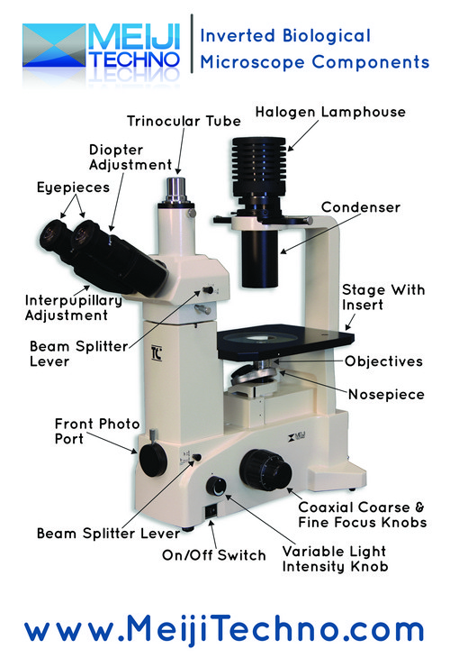
TC-5400L Binocular Inverted Brightfield/Phase Contrast Biological ...
Scanning Electron Microscope (SEM) - Diagram, Working … Mar 23, 2022 · Definition. Scanning electron microscope is a classification of electron microscope that uses raster scanning to produce the images of a specimen by scanning using a focused electron beam on the surface of the specimen.. An SEM creates magnified images of the specimen by probing along a rectangular area of the specimen with a focused electron beam.

Euglena Acus 2 - BF microscope 1250x - YouTube
Compound Microscope Parts, Function, & Diagram | What is a Compound ... The definition of a compound microscope is "an upright microscope that utilizes two different lenses to magnify the size of the objects being viewed." The name itself describes what it is. The tern...
Understanding the Compound Microscope Parts and its Functions
Compound Microscope | Definition, Examples, Diagrams - Toppr Compound microscope is an optical device used to obtain very large values of magnification. It is used to see microscopic objects like microorganisms. It comprises of two convex lenses and magnification occurs in both of them. Its basic components are: 1. Objective: Convex lens placed near the object. Object is placed just beyond the focal ...

Microscope With Labels Clip Art at Clker.com - vector clip art online ...
Parts of a Compound Microscope and Their Functions Compound microscope mechanical parts (Microscope Diagram: 2) include base or foot, pillar, arm, inclination joint, stage, clips, diaphragm, body tube, nose piece, coarse adjustment knob and fine adjustment knob. Base: It's the horseshoe-shaped base structure of microscope. All of the other components of the compound microscope are supported ...
Post a Comment for "44 compound microscope diagram"